Live images from inside materials
Erlangen, Germany, December 2, 2014: X-rays are a tried and tested way to investigate components and materials. Researchers are now developing an X-ray detector capable of delivering particularly high-quality 3D images in real time. This will make it possible to precisely reconstruct even the processes going on inside materials and e.g. provide a reliable way of detecting minoscule faults.
In medicine, X-rays provide high-resolution images of our insides to help doctors make a definitive diagnosis. Industry uses X-rays, too – as a reliable, non-destructive way of seeing what’s hidden on inside materials and components and to check for cracks or irregularities. However industry additionally draws upon different technologies that are not used in the medical field. Whereas medical X-ray machines have been specifically designed for human test subjects, industrial X-ray machines are used to analyze objects that vary much more in their size and material composition. This calls for X-ray equipment that is correspondingly more flexible.
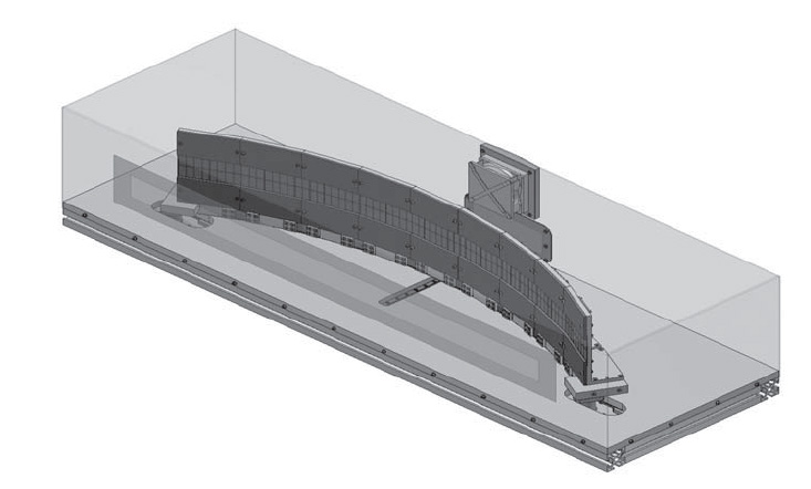
Researchers at the Development Center for X-Ray Technology EZRT, a division of the Fraunhofer Institute for Integrated Circuits IIS, have developed MULIX – an X-ray detector for industrial computed tomography (CT) based on the design of medical X-ray devices. “Our challenge was to combine high image quality with a high degree of flexibility,” explains Frank Nachtrab from the EZRT. MULIX harnesses two concepts already in use, making it a kind of hybrid solution that incorporates elements of line and flat-panel detectors commonly used in industry. The researchers already know what they plan to achieve with their work: “We’ve had very promising results with our demonstrator and have shown that MULIX works. We’re now looking for industrial partners to help develop a MULIX prototype,” says Nachtrab.
Combining the benefits of two different methods
Single-line detectors use a fan-shaped beam to X-ray a certain section of the test object, whereas flat-panel detectors are working with a cone-shaped beam that encompasses the entire object. There are pros and cons to both solutions. A flat-panel detector will quickly give you a 2D image of the entire object. However, it causes scattered radiation – in other words, rays deflected by the test object – which greatly impairs image quality. A single-line detector is less sensitive for scatter and will therefore deliver extremely sharp images. But since it captures only a small portion of the test object, this scanning method is much more time-consuming. “We have combined the benefits of the two solutions,” says Nachtrab. The new equipment is based on a detector with multiple lines, a design that until now has been used only in the medical field. Multipleline detectors work according to the same principle as their single-line counterparts, but can also cover larger areas and thus radically reduce the scanning time. MULIX uses a total of 256 lines, allowing it to scan larger objects such as car body parts very quickly. What’s really remarkable is that the new detector delivers images so fast, it becomes possible to use CT techniques to make a 3D-scan of the object almost in real time.
MULIX opens up new application opportunities in materials research and quality assurance, which would allow the automotive industry, aerospace and research institutions to observe processes going on within the materials they use. “When testing mechanicalb properties such as tensile strength, we can use the images we get to see just how a compromising fault comes about,” says Nachtrab. The researchers also came up with an innovative solution for the detector’s mechanics: “This enhances the quality of the images further,” says Nachtrab. Unlike commercially available detectors, it is possible to adjust MULIX’s curvature. This ensures the flexibility that industrial CT needs to adapt the system to the various sizes and material properties of test objects.
